


Convex
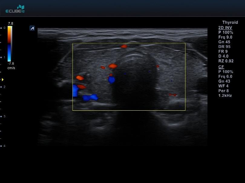
Linear
Phased array
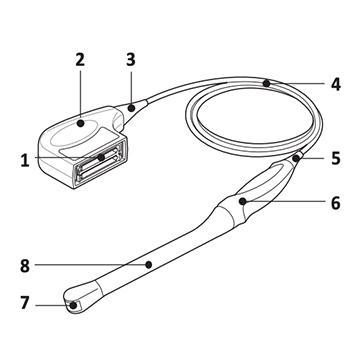
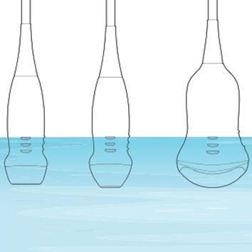
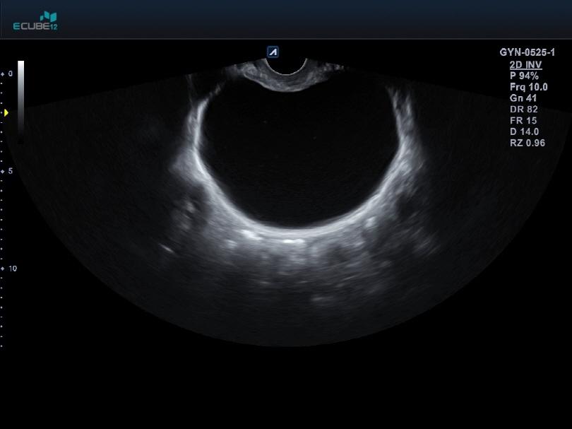
Endocavity
Volume
Others
Convex


Linear


Phased array


Endocavity


Volume


Others