


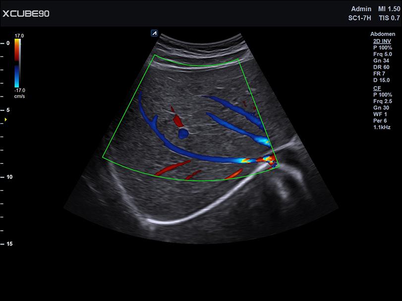
Spatial Resolution
Frame rate
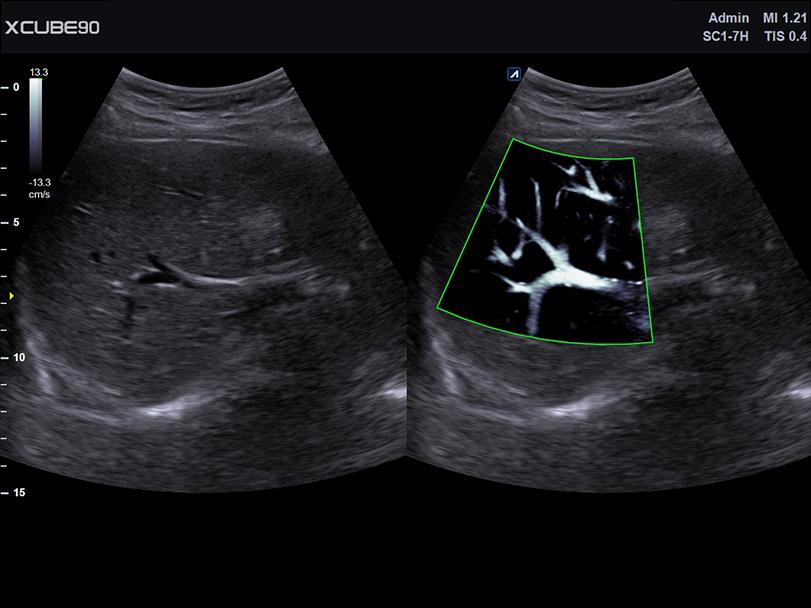
Sensitivity & Penetration


General imaging
MSK
Women’s Health
Cardio vascular
General imaging


MSK


Women’s Health


Cardio vascular


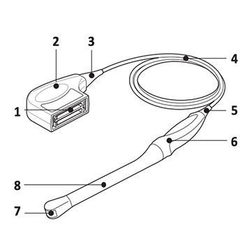
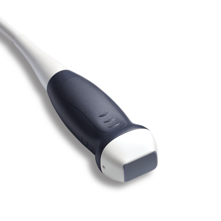
Convex
Linear
Phased array
Endocavity
Volume
Others
Convex


Linear


Phased array

Endocavity


Volume


Others