
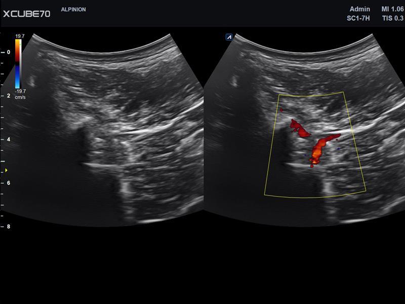
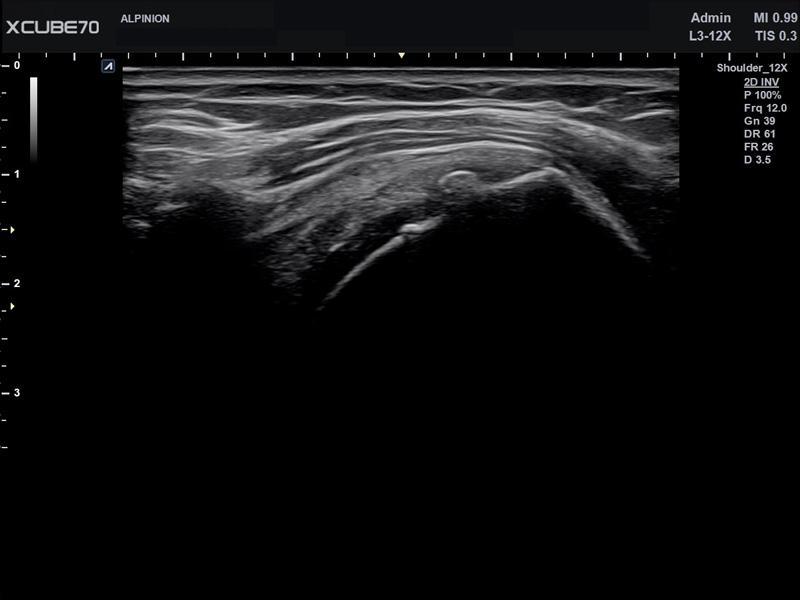
Spatial Resolution
Frame rate
Sensitivity & Penetration


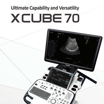
Enjoy a larger widescreen and clear, high definition images.
12.1 inch high resolution touch panel allows users to
tilt up to 15° for user convenience.

Providing more ease to pick up
the transducer and gel, the additional
holder provides a convenient diagnostic environment to fit the body shape of user.
The control panel may be easily adjusted vertically with its motorized button.
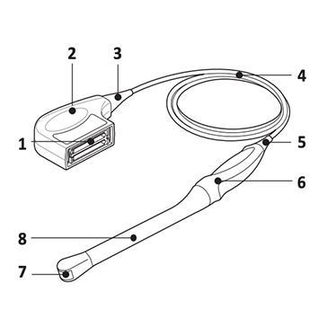
5 transducer connectors are composed of 4 active and 1 optional.

Providing more ease to pick up
the transducer and gel, the additional
holder provides a convenient diagnostic environment to fit the body shape of user.


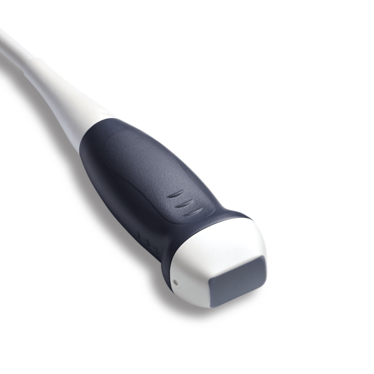
Convex
Linear
Phased array
Endocavity
Volume
Others
Convex


Linear


Phased array
Endocavity


Volume


Others