Innovation Meets Portability. Indeed.
Offering a powerful and stable cart-based system architecture and breakthrough design with sliding keyboard cover,
The E-CUBE i7 delivers excellent imaging performance with an ergonomic design for your comfort during scanning.
The compact i7 is versatile, featuring application-specific imaging technology with advanced transducers.
From the bedside to the procedure room, the E-CUBE i7 provides ease-of-use and workflow efficiency.
The innovative system architecture
The advantages of a cart-based system made the innovative portable system, E-CUBE i7.
Thanks to this powerful and stable architecture,
the E-CUBE i7 delivers remarkably excellent and uniform image performance throughout its lifetime.
Clinical Image


Excellent Imaging Quality
Crystal Signature™
-
High class single crystal transducer technology
-
ALPINION’s world leading single crystal technology, provides higher sensitivity, better penetration, and less heat degradation.
High frequency enhanced linear transducer
-
The E-CUBE i7 features a high-frequency linear transducer, the hockey stick-shaped IO8-17T.
-
The IO8-17T is ideal for imaging for superficial, nerve blocks, and vascular procedures.
Optimal Clinical Environments
-
Experience the sliding keyboard cover. Enjoy the comfort.
-
More than 80% of sonographers experience work-related pain and more than 20% of these suffer a career-ending injury.*
-
* Society of Diagnostic Medical Sonography Industry Standard for the Prevention of Musculoskeletal Disorders in Sonography. May 2003.

Mobility
1 hour battery life / 7.2kg Lightweight system

Fast boot-up
Less then 45 seconds

Library Quiet
Library(30dB) / E-CUBE i7(31dB)
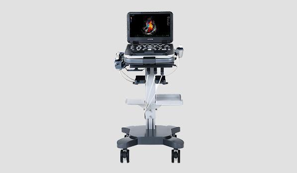
Extended transducer
Connection up to 3 ports
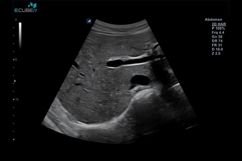
White Paper
-
A Study of Wrist Angles when using portable ultrasound system with a wrist supporting device
-
Tae-Hoon Lee, Suh-Wo Choi, Hak-Jun Kim Department of Orthopedic Surgery, Korea University Guro Hospical, South Korea
Software Technologies
-
Needle Vision™
-
Panoramic Imaging
-
Xpeed ™ (Auto Image Optimization)
-
Stress Echo
-
Auto IMT measurement
-
3D4D
-
CW(Continuous Wave)
Transducers
C1-6T
Convex transducer (1-6MHz)
Application
Abdomen, Renal, Fetal Echo, EM Gynecology O bstetrics MSK, Urology
C5-8NT
Microconvex (5-8MHz)
Application
Abdomen, EM, Cardiac, Pediatric
L3-12HWD
High density wide footprint linear transducer (3-12MHz)
Application
Breast, EM, MSK, Vascular, Small Parts, Appendix
L3-12T
Linear transducer (3-12MHz)
Application
Breast, EM, MSK, Vascular, Small Parts, Appendix
IO3-12
Intraoral transducer (3-12MHz)
Application
EM , Small Parts
IO8-17T
High frequency hockey stick (8-17MHz)
Application
Small Parts, MSK
SP1-5T
Single crystal Phased array transducer (1-5MHz)
Application
Abdomen, Cardiology, EM, TCD
SP3-8T
Single crystal phased array transducer (3-8MHz)
Application
Abdomen, Cardiac, EM, Pediatric
EV3-10T
Endocavity transducer (3-10MHz)
Application
GYN, OB, Fetal Echo, Urology, EM
EC3-10T
Endocavity transducer (3-10MHz)
Application
GYN, OB, Fetal Echo, Urology, EM
VC1-6T
Volume Convex (1-6MHz)
Application
Abdomen, OB, GYN, EM
CW5.0
Pencil type transducer (5.0MHz)
Application
Cardiac
CW2.0
Pencil type transducer (2.0MHz)
Application
Cardiac
Resources
Tutorials
Step 1. OVERVIEW
Step 2. USER SETTING
Step 3. TRANSDUCER
Step 4. BEGINNING AND EXAM
Step 5. IMAGING FUNCTIONS
Step 6. OPTIMIZE IMAGE
Step 7. MEASUREMENT AND REPORT